The primary aim for the establishment of the Department of Anatomical Pathology was to train pathologists in the Taipei City government hospitals. Since that time, 8 pathologists have been trained and licensed and are currently working in various well-known hospitals, thus fulfilling our initial objective. Our current focus is on providing quality diagnostic services for small-to-medium sized hospitals and clinics. In recent years, the institute has also set up and developed the Molecular Pathology Diagnosis Lab, earning it a national recognition as a “Benchmark Laboratory,” for its advanced diagnostic methods and comprehensive clinical services. The Department of Anatomical Pathology is currently one of Taiwan’s biggest Pap smear screening centers. The members of this strong work force currently include 6 pathologists, 12 cytotechnologists, 8 medical technologists, 2 laboratory technicians, 6 clerical staff, and 1 specimen collection staff members.
The main functions are as follows
Surgical pathology
1. Pathology biopsy diagnosis2. Frozen biopsy diagnosis3. Special staining4. Immunohistochemistry staining
5. Paraffin tissue block and paraffin section production
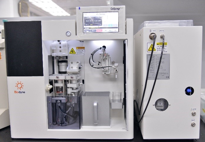
Cellprep PLUS
Cytological pathology
(1) Cervical smear: The Pap smear examination The Pap smear examination is the simplest and most effective way for early stage detection of cervical cancer. Since the establishment of the National Health Insurance Program in 1995, females aged 30 and above have been entitled to free annual Pap smear examinations. Our institute is recognized by the Department of Health (now Ministry of Health and Welfare) as a certified pathology medical institution, that registers reports directly with the government’s screening statistics program.
(2) Liquid Thin Prep Cytology This procedure is different from the traditional Pap smear in which the collected cervical brushings specimen is placed into a vial containing a special preservative, instead of being directly smeared on top of a slide. The sample is then made into a thin smear by an automated smear- making instrument.
(3) Non-gynecological Cancer Screening An Exfoliative Cytology examination includes 2 other categories apart from the Pap smear. The first is the body fluid cytology exam, which includes testing of sputum, urine, ascitic fluid, cerebrospinal fluid, pleural effusion, pericardial fluid, bronchial brushing, and bronchial washing. The second is the aspiration cytology examination, which includes the thyroid, lymph node, breast, lung, liver, pancreas, and tumor aspirations.
(4) Human Papilloma virus Screening After 50 years of relentless research, it has been confirmed that cervical cancer is definitely linked to the Human Papilloma virus (HPV) infection. The HPV genome is a double-stranded DNA with more than 100 genotypes of which 30~40 are sexually transmitted. Thirteen of them, namely Types 16, 18, 31, 33, 35, 39, 45, 51, 52, 56, 58, 59, and 68 are considered “high risk” types related to cancer. HPV screening allows early detection and early treatment if necessary.
Molecular Pathology
(1) Anaplastic lymphoma kinase (ALK) Protein for lung cancer detection
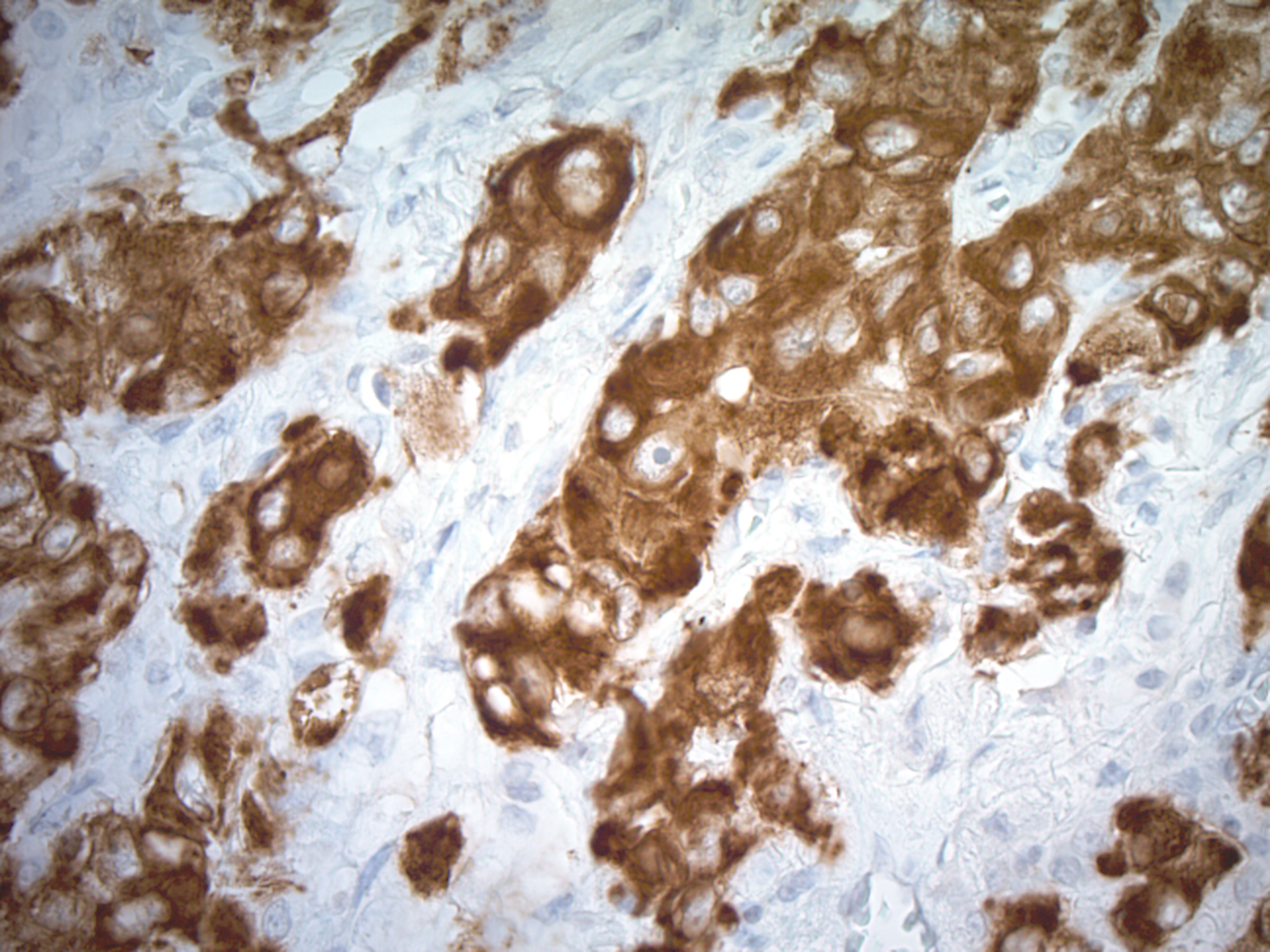
ALK |
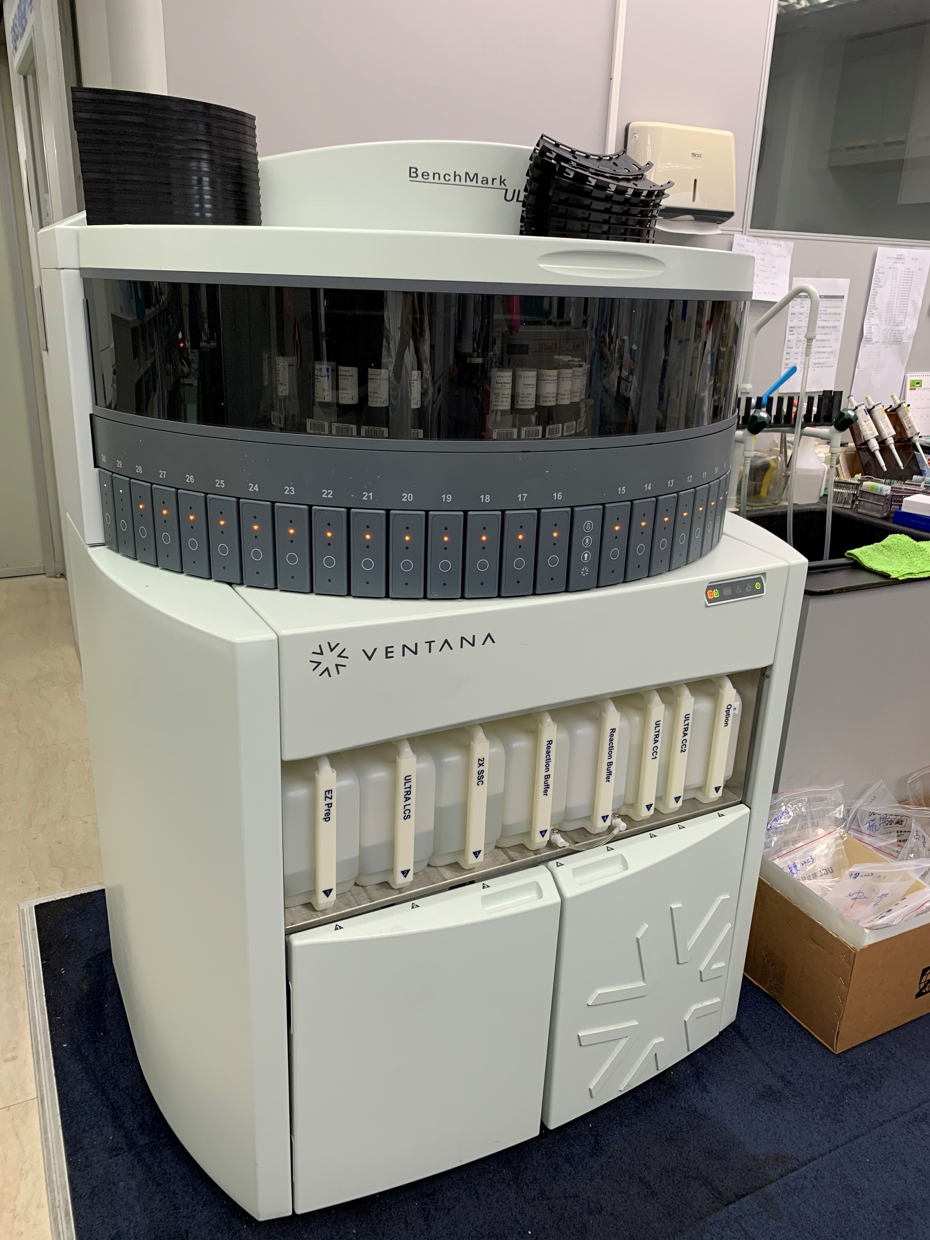
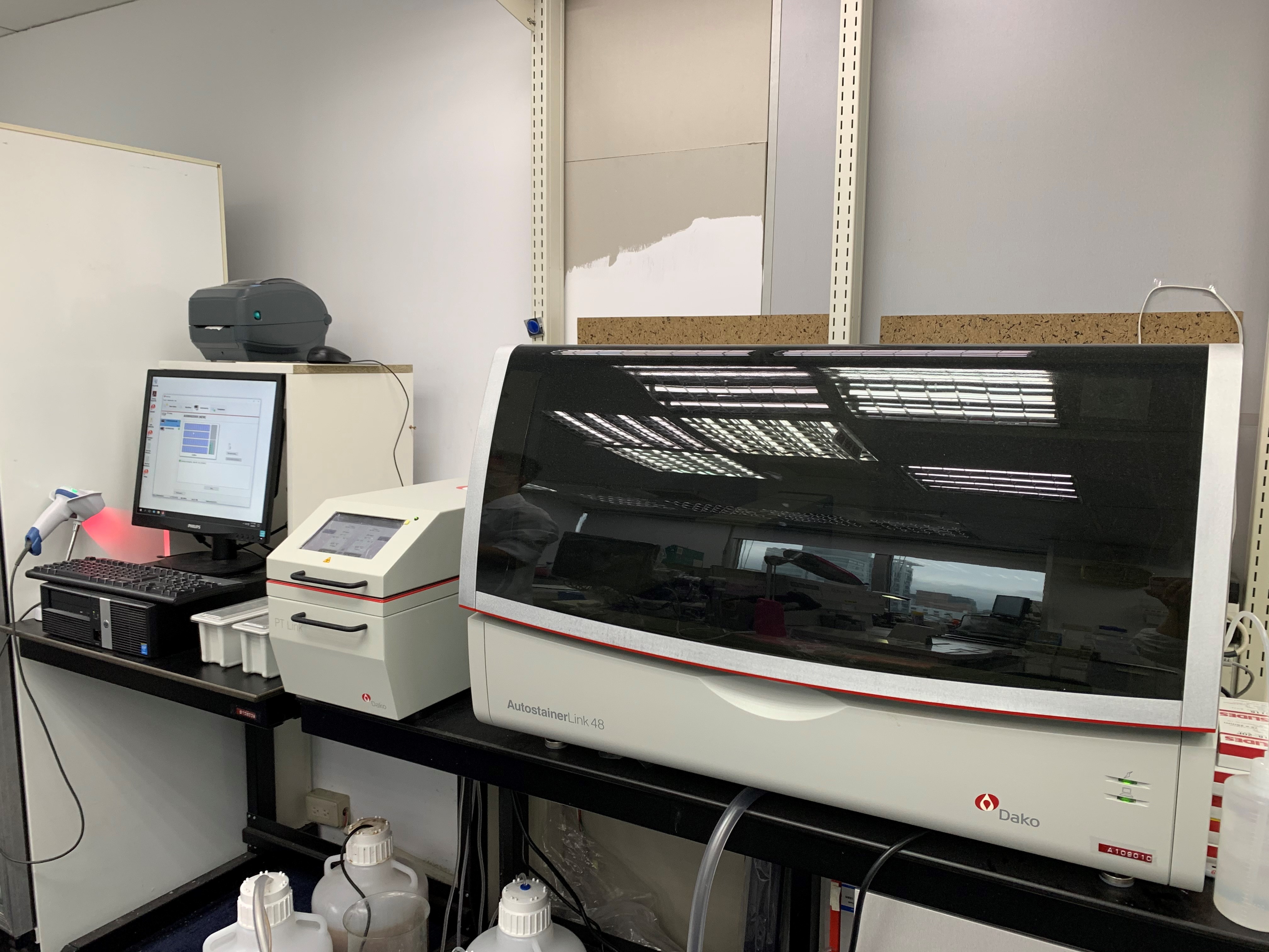
The OptiView DAB IHC Detection Kit and automated immunohistochemical staining with automatic systems, primarily uses formalin-fixed and paraffin-embedded non-small cell lung cancer tissue specimens. The anti- ALK (D5F3) rabbit monoclonal antibody is the primary monoclonal antibody that combines ALK and specimens. By using the OptiView DAB IHC detection Unit and OptiView amplification kit, this specific antibody can be visually observed under an optical microscope. This test assists in screening suitable candidates for XALKORI (crizotinib) treatment. |
(2) HER2/neu gene amplification for breast cancer detection
The HER2 Dual ISH DNA Probe Cocktail was designed to quantitatively detect amplification by microscopy; the HER2 gene, via two colour chromogenic in situ hybridization(ISH) in formalin-fixed, paraffin-embedded human breasts; and gastric cancer, including the gastroesophageal junction. Following staining on automated slide strainers, the result can be used as an aid in the assessment of patients for whom Herceptin (trastuzumab) treatment is being considered.
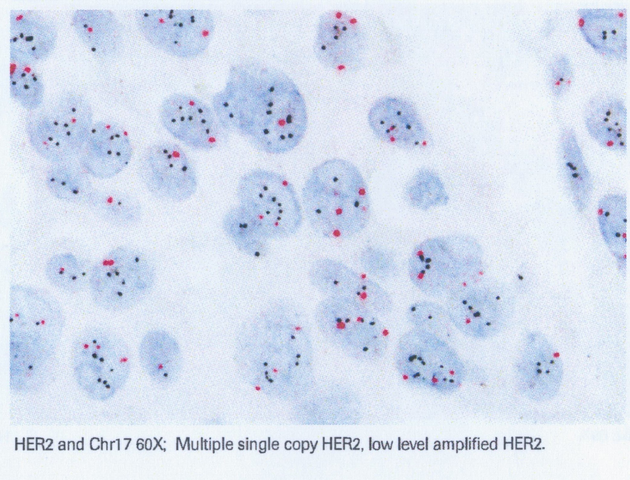
|
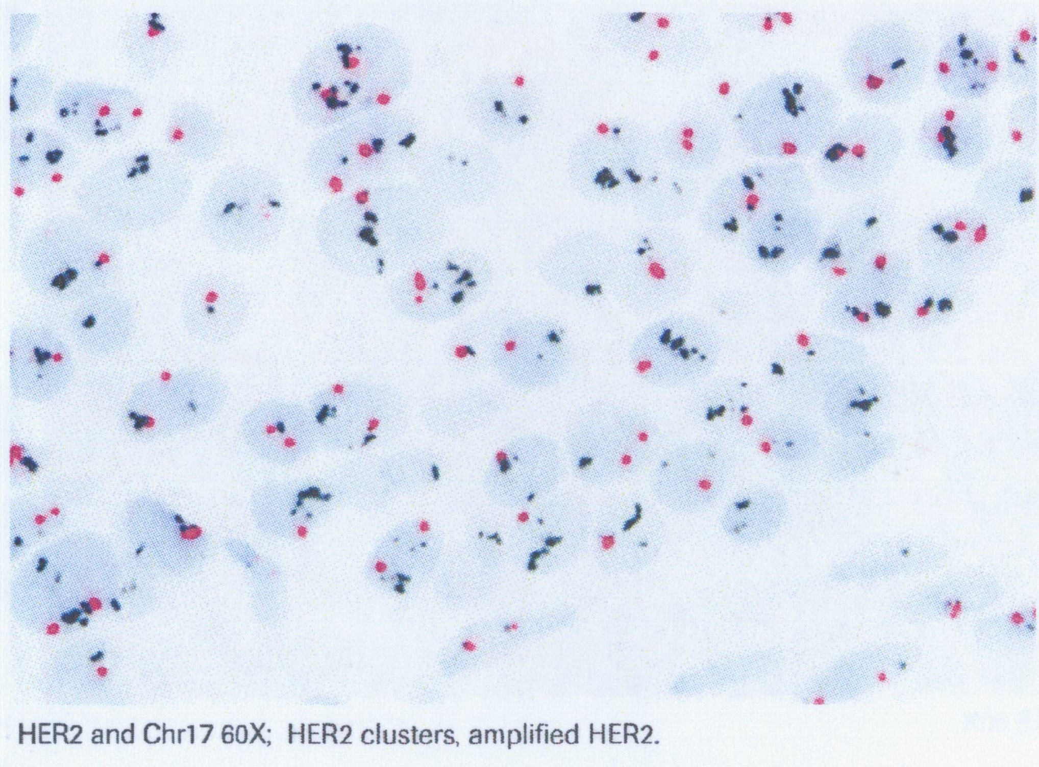
|
| DISH | |
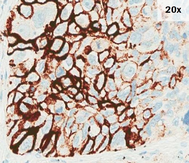
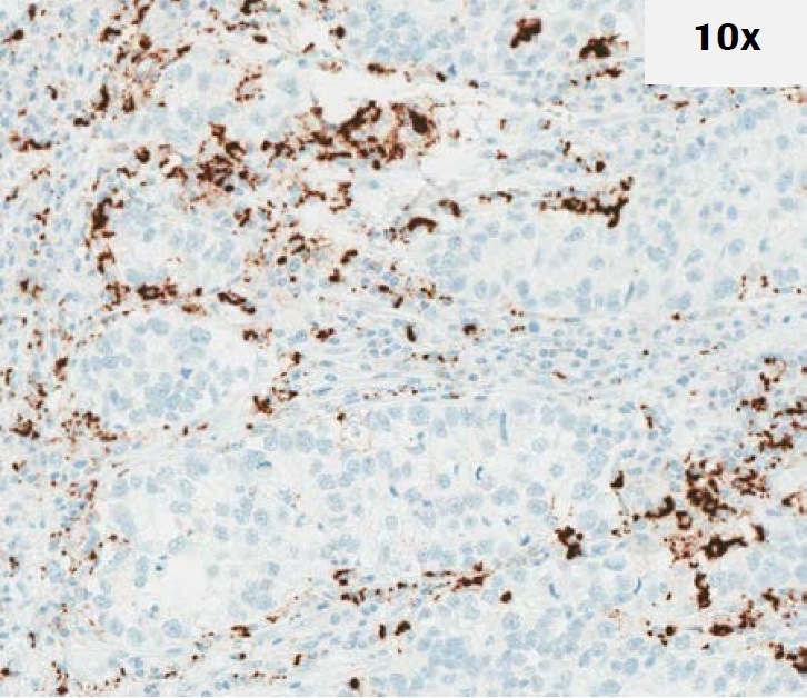
(3) PD-L1
PD-L1 is an immune-related biomarker that can express in the tumor cells. The patients’ eligibility for anti-cancer immunotherapy highly relates to the expression of the PD-L1 protein in the tumor. The primary detection method for the PD-L1 protein in non-small cell lung cancer (NSCLC) is immunohistochemical stains. With the automatic immunohistochemical staining system, a variety of PD-L1 antibody clones produced by different manufacturers could be applied on the formalin-fixed and paraffin-embedded tissue specimen. The staining results aid the clinicians in the decision on whether or which anti-cancer immunotherapy they should prescribe for the patient.
|
|
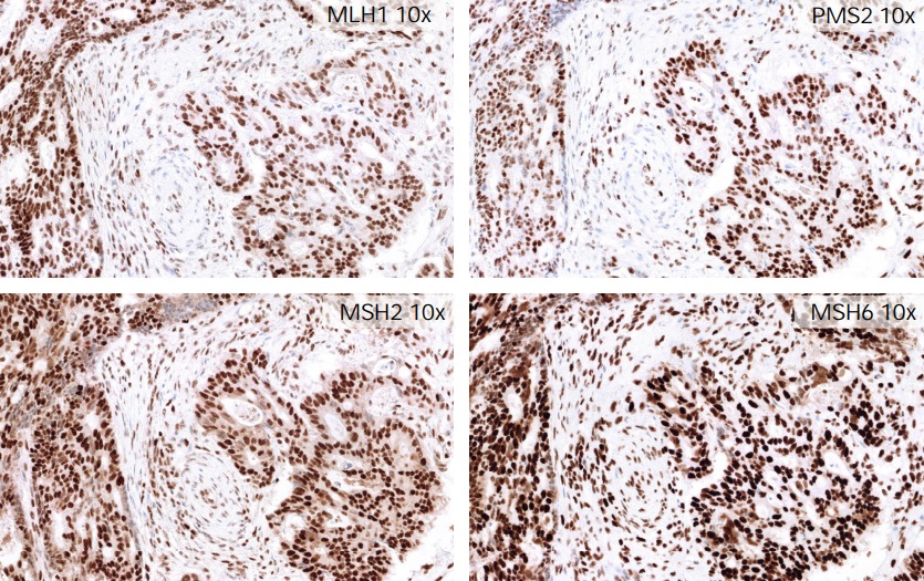
(4) Mismatch repair gene (MMR gene) for colorectal cancer detection
Hereditary nonpolyposis colorectal cancer (HNPCC) is one of the most common diseases of hereditary colorectal cancer, accounting for about 2-5% of all colorectal cancers. It is caused by the mutation of mismatch repair gene (MMR gene). These gene products are mainly related to gene repair in cells, including: MLH1, MSH2, MSH6, and PMS2. These four gene mutations that induces HNPCC production is confirmed. Therefore, IHC is used to detect the protein expression of MMR gene in cancer tissue. If there is no expression, it means that the relative gene has mutated and loses its function. intraepithelial lesion), or in patients with positive high-risk HPV test results.
|
| MMR |
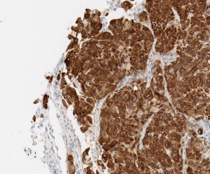
(5) ROS1 protein for lung cancer detection
ROS1 is a tyrosine kinase receptor in the insulin receptor family. In non-small cell lung cancer, multiple fusion partners of ROS1 genes have been confirmed. These fusion proteins have kinase activity, which can enhance cell proliferation and reduce cell apoptosis.This test assists in screening suitable candidates for XALKORI (crizotinib) treatment.
|
| ROS1 |
Our Depar tment al so provide s par af f in making and embedding tissue services to various universities and research labs, not only for diagnostic purposes, but also with the goal of supporting the mission to help these institutions improve the quality of their biopsy interpretation skills. Our services includes generating human and animal tissue biopsies, hematox ylin- eosin (HE) staining, immunohistochemical staining, frozen tissue biopsies, and various special stains such as Masson- trichrome and PAS, etc.
|
|
| VENTANA BenchMark® ULTRA | Dako Autostainer Link48 |
Items of Examination
Surgical Pathology
Surgical biopsy
Frozen section
Immunohistochemistry staining
EGFR
ALK
MMR
PD-L1 (22C3、SP263、28-8、SP142)
ROS1
HER2 Dual ISH
Paraffin-embedding tissue service
Cytological Pathology
Cervical smear
Liquid based thin smear
Non-gynecological body fluid cytology
Non-gynecological needle aspiration cytology
Human papilloma virus DNA test

